When is voice surgery indicated for vocal cord leukoplakia?
The indication for surgery of vocal cord leukoplakia is always given when anti-inflammatory therapy lasting approximately 10 days does not resolve the whitish patches. Under all circumstances, the tissue from the leukoplakial area should be tested histologically. That is why the patients need to be informed that they require phonosurgery for reasons of safety. Otherwise, a potentially malignant vocal cord leukoplakia could develop into a laryngeal cancer.
Occasionally, patients with chronic laryngitis will suffer repeated bouts of leukoplakia lesions on their vocal folds. Here, control biopsies of the findings are recommended at regular intervals. Important note: just because the histological finding was normal in the past does not necessarily imply that the next histological finding will likewise still be normal in the future. Any newly emergent case of vocal cord leukoplakia must be re-evaluated based on the most recent findings.
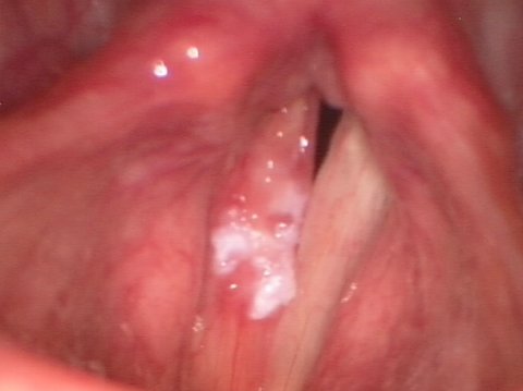
Vocal cord leukoplakia before surgery

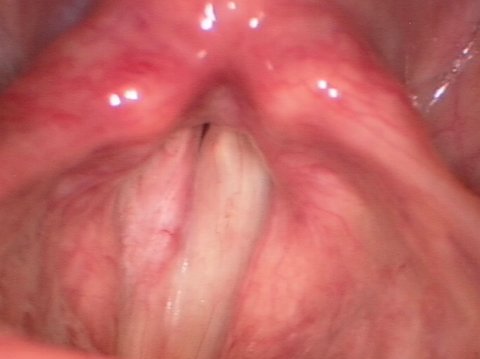
Vocal cord leukoplakia after surgery


How is vocal cord leukoplakia managed surgically?
Vocal cord leukoplakia is a condition primarily affecting the superficial mucous membrane of the vocal folds. However, it can also grow into the deeper layers of the vocal fold mucosa.
If the voice surgeon now aims to remove a potentially malignant finding with a safety margin while at the same time preserving the patient’s voice in generally good condition, a surgical method must be selected which allows the leukoplakia to be separated and removed from the healthy vocal fold tissue without any major tissue loss.
Removal of a large area of leukoplakial vocal cord tissue from healthy tissue would mean sacrificing mucosal layers capable of vibration in order to be on the safe side. Such an excision is usually accompanied by a significant, often irreversible worsening of voice quality. Such outcomes are repeatedly observed after laser surgeries.
For this diagnosis of vocal cord leukoplakia, the method of "hydrodissection” has proven its merits in that it preserves as much tissue as possible when the whitish patches need to be lifted off. This method involves using a fine, angled needle through which saline solution with an added hemostatic is injected under the leukoplakia lesion. The procedure elevates the lesion up off the healthy vocal fold tissue to permit its microsurgical removal. That way, the smallest volume of healthy tissue is taken up during the procedure.
However, if the leukoplakial lesion has already grown deep into tissue layers of the vocal fold, hydrodissection is no longer possible. In such cases, extensive tissue excision is almost always necessary to successfully manage the process of invasive proliferation.
Vocal cord leukoplakia before surgery
Vocal cord leukoplakia after surgery

What happens during the postoperative phase after voice surgery for vocal cord leukoplakia?
After voice surgery for vocal cord leukoplakia, the patient should maintain an approximately 10-day period of vocal rest. During this time, the wound is allowed to heal while reepithelialization takes place. Reepithelialization is the natural process of a fine layer of new skin (epithelium) growing over the surface of the wound.
It is imperative that voice rest be maintained during this period in order to prevent wound healing disorders and adhesions or scarring. Additionally, the patient must abstain from smoking cigarettes and drinking alcohol during this period due to the associated risk of bleeding.
This period of vocal rest should be followed by intensified voice therapy with a highly qualified therapist who is specialized in voice disorders.
In most cases, the described method of plastic reconstructive surgery is successful in removing superficial vocal cord leukoplakia while preserving the patient's good voice quality. Consequently, voice therapeutic interventions also usually yield satisfactory outcomes in terms of voice quality.

Vocal Fold Augmentation in Patients with Glottic Insufficiency (After Cancer Surgery)
When extensive tumor involvement of the vocal folds mandates extensive tissue excision, it can be expected that a significant voice disorder will be the result. Absent their mucosal layers capable of vibration, the vocal folds lose their ability to close completely during phonation. This produces a very breathy, almost toneless voice.
If, in such a case, voice therapeutic exercises do not deliver the desired success, voice improvement surgery may become necessary. One available option is augmentation of the diseased vocal fold with autologous fat tissue. The phonosurgical options for this indication are described in detail under Voice Surgery for Glottic Insufficiency After Cancer Surgery.


