What are vocal cord leukoplakia and laryngeal cancer?
Laryngeal Cancer (Laryngeal Carcinoma)
Laryngeal cancer ranks among the most common malignant tumors observed in otolaryngological practice and occurs in men with a much greater frequency than in women; the age peak is between 55 and 65 years of age, respectively.
Laryngeal carcinomas are subdivided into three groups according to their localization.
- Glottic cancer (carcinoma of the vocal folds) 60%
- Supraglottic cancer (carcinoma above the vocal fold level) 40%
- Subglottic cancer (carcinoma below the vocal fold level) 1%
Among all laryngeal cancers, glottic cancer, which is primarily limited to the level of the vocal fold, occurs with a prevalence of up to 60%.
Since Dr. Wohlt exclusively operates on laryngeal carcinomas of the vocal folds, the following passages aim to address cancer of the vocal cords, in other words, glottic laryngeal cancer only.
Squamous cell carcinoma of the vocal fold

Vocal Cord Leukoplakia
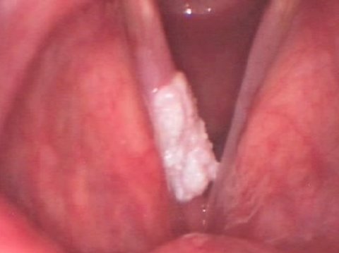
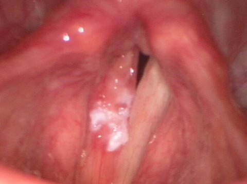
Leukoplakia presents as a whitish, plaque-like tissue lesion on the vocal fold surface that can exhibit circumscribed as well as diffuse spread.
Vocal cord leukoplakia can develop into vocal fold cancer where external visual inspection does not allow exclusion of malignancy with certainty. That is why histological tests by the pathologist are required in such cases.
Vocal Cord Leukoplakia


How do vocal cord leukoplakia and laryngeal cancer develop?
The most frequent causes that promote the development of vocal cord leukoplakia and laryngeal cancer are smoking cigarettes and drinking alcohol. The prior medical history of afflicted patients will most overwhelmingly list clear signs of at least one of these two risk factors.
In addition, laryngopharyngeal reflux, i.e. the backflow of gastric acid into the laryngeal and pharyngeal region is presumed to promote the development of vocal cord leukoplakia and laryngeal cancer.
Leukoplakia on the vocal cord can also develop as a result of chronic laryngitis. In patients suffering from years of persistent chronic laryngitis mostly due to habitual cigarette smoking, leukoplakia can primarily develop and then secondarily lead to vocal fold cancer.

What symptoms are caused by vocal cord leukoplakia – laryngeal cancer?
Vocal cord leukoplakia, which is to be regarded as a premalignant lesion to laryngeal carcinoma, usually causes hoarseness the severity of which is proportionate with the extent of the disease. Hoarseness is the cardinal symptom noticeable in mucosal diseases of the vocal folds. When the lesion grows to a significant size, the patients may have a permanent feeling of a lump in their throat and experience difficulty when swallowing.
It should be specifically pointed out here, however, that the symptom of hoarseness alone is not a conclusive indication for a benign or malignant process of the vocal folds. Therefore, any hoarseness persisting longer than two to three weeks must urgently be examined by an ENT specialist using laryngoscopy.

What treatment options are available for vocal cord leukoplakia?
- Voice surgery
When the whitish circumscribed plaques appear for the first time:
- Pharmacotherapy
- Inhalation
Because vocal cord leukoplakia could be a precursor to laryngeal cancer, a histological test should be conducted under all circumstances to rule out any malignancy. For that reason, a phonosurgical intervention is the method of choice.
However, intensive anti-inflammatory treatment with medication and inhalation is allowed for approximately 10 days to try to treat newly emergent whitish circumscribed plaques.
If this therapy proves to have no effect on the plaques, a surgical Intervention should be undertaken immediately.
Phonosurgery for vocal cord leukoplakia should always take a voice preserving-approach. If the removal of leukoplakia from healthy tissue is too generous, significant and irreversible hoarseness is frequently the postoperative outcome. Therefore, a conservative surgical method should be selected to treat these cases. The phonosurgical options for this intervention are described in detail in the section Voice Surgery for Vocal Cord Leukoplakia.
Example: Phonosurgical management of vocal cord leukoplakia
Vocal cord leukoplakia before surgery
Vocal cord leukoplakia after surgery
Example: Phonosurgical management of vocal cord cancer
Vocal cord cancer before surgery
Vocal cord cancer after surgery

Truths and Myths About Vocal Cord Leukoplakia
Vocal cord leukoplakia is by definition a premalignant lesion, in other words, a potential precursor to laryngeal cancer. Nevertheless, its visual appearance is not sufficient to rule out malignancy or confirm the benign nature of the lesion. Therefore, the patients should be advised most emphatically to undergo a phonosurgical intervention to treat these types of lesions when they prove unresponsive to drug or inhalative treatment. One of the primary aims is to take a biopsy of the tissue for histological examination.
If histology confirms malignancy, the further therapeutic strategy must be discussed. Voice surgery, irradiation and chemotherapy are the options under consideration.
On the other hand, it should be noted that whitish changes to the vocal folds will frequently respond very well to anti-inflammatory drugs and tend to resolve under this medical therapy.

Case Report: Vocal Cord Leukoplakia
Over a period of several months, this 52-year-old music teacher had been noticing increasing hoarseness. He reported initially having only a slightly veiled rough voice. Over the course of months, however, severe hoarseness had set in that was additionally remarkable because his voice was abnormally deep. Singing during music lessons cost him increasing effort and he was recently unable to reach a high tessitura pitch.
The patient reported smoking around 10 cigarettes a day. He only drank alcohol rarely. He remembered that he had suffered laryngitis around six months prior that caused him considerable impairment for months. This was when the patient experienced the onset of his hoarseness.
The videostroboscopic examination showed a marked leukoplakial change in the mid-third of the right vocal fold. The lesion was no longer limited to a circumscribed area, but already exhibited what is called exophytic growth, in other words, a diffuse proliferation of tissue on the surface of the vocal fold. Compared to the left, the right vocal fold looked much more inflamed although the respiratory mobility of the vocal folds was normal. The vibrations of the right vocal fold were likewise restricted in comparison with the left. His voice was hoarse and his average speaking pitch was markedly lowered. His singing voice had essentially disappeared.
The patient was urgently advised to undergo a phonosurgical procedure. This procedure relied on the method of plastic reconstruction. Intraoperatively, the leukoplakial area was found to have spread widely over half of the vocal fold.
Therefore, the method of gentle decortication was selected. Most of the superficial mucous membrane was removed during this procedure. The deeper layers of the lamina propria were preserved so that the patient's postoperative voice could be expected to remain mostly good.
The histological findings proved the lesion indeed to be a precursor to laryngeal carcinoma. It was what is called a carcinoma in situ. This has to be regarded as premalignant because the tissue had the potential to develop into laryngeal cancer if it had remained in the patient.
After a 14-day period of vocal rest and supportive drug therapy, the patient's voice had returned with a good quality. His voice quality was resonant and clear. The patient reported his average speaking pitch as mostly normal. The patient himself perceived his own voice as pleasant and functional.
After another six months, the patient returned for a follow-up examination. He continued to work as a music teacher. His voice was mostly normal. During the videostroboscopic examination, smooth, bland vocal folds were seen, both demonstrating a good vibratory patterns. Compared to the not operated one, the operated vocal fold only exhibited a minor limitation in the mucosal wave.


